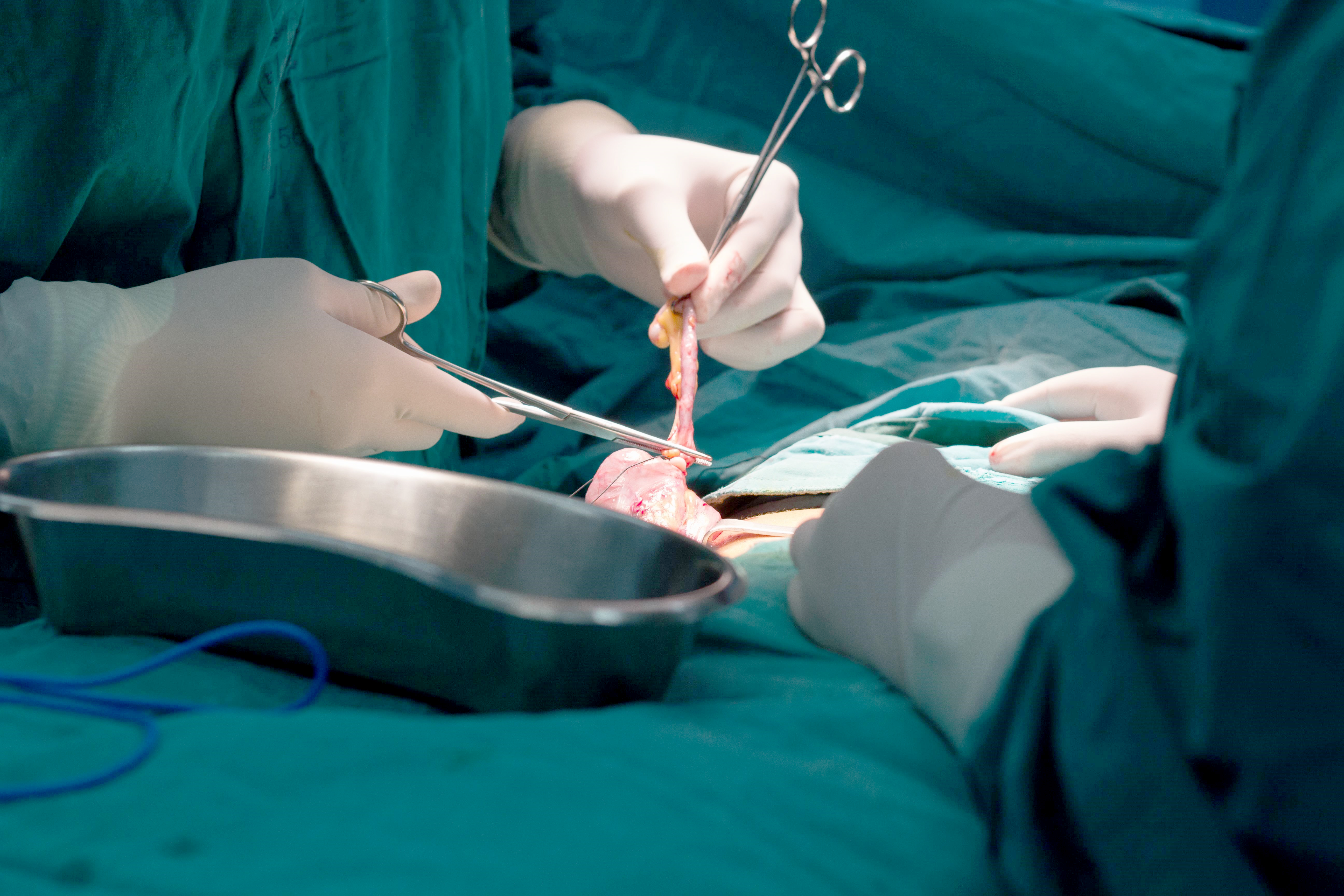
Appendicitis
I’ve written before about appendicitis and the research going on to find the best type of treatment. A big part of that research is to make double-dog sure that it keeps people safe because this thing used to kill people right and left!
Contrary to what the charlatans on the internet try to get you to believe, NOT EVERYONE is safe waiting to see if antibiotics are the only treatment needed; and, not every doctor is bad just because they recommend surgery.
Vintage painting of Hippocrates examining a sick boy with appendicitis
I know, in my previous article I discussed that some clinicians were discovering that in some cases patients recovered from appendicitis with only antibiotics when surgery was delayed or unavailable for some reason; and that we wanted to see how or if that applied to a safe “standard of care” treatment algorithm.
Afterwards, I noticed that many other “lay-person” blogs and news reporters began sensationalizing this as a “new treatment” or “major breakthrough” while the truth is that: it’s an old idea which is now being tested for safety and most likely will NOT apply universally to ALL patients with abdominal pain that might be appendicitis.
Let’s maintain some intelligent reasoning here:
Appendicitis: Treatment Options
Antibiotics, surgery or blood-letting
First, this problem has been around since Hippocrates and killed a lot of people. Second, surgical treatment was an option long before antibiotics were recognized. Third, the abdomen contains a lot of stuff that can hurt besides the appendix; and, Fourth, the appendix is not located in the same place in everyone!
That’s a lot of variables and add to that the huge number of bacteria which cause it, the growing antibiotic resistance of those organisms, the use of laparoscopic techniques these days, the wide variation in symptom tolerance and descriptions between patients AND the sheer timing of seeking help—well, it means NO TWO PATIENTS ARE THE SAME.

And, keep in mind, the only reason a sensible person would even dare do something that risks a patient’s life or well being like this is because we now have imaging technology which can give us an inkling about what’s going on inside the patient’s belly without opening them up. There is still no absolute fool-proof diagnostic test to make the diagnosis of appendicitis except by looking directly at it!
So, IF there’s no complications, IF imaging is available, IF other laboratory tests are ok, IF antibiotics are available and working, IF the patient is being carefully observed in the hospital to be kept safe and IF surgery is immediately available should things “go south” THEN a doctor might be daring enough to use antibiotics as the sole treatment.
Then, you’ve got to consider: what do we do next? Will it come back? If so will it be harder to diagnosis or treat?
Appendectomy Facts
Let’s discuss a few of the other facts we’ve learned while we’ve been studying the issue. You already know, of course, that the appendix is a small tube-like piece of tissue at the end of the small intestine full of lymphatic tissue sort of like the tonsils—we still don’t really know exactly what it’s for.
It can get plugged by feces, foreign material, a follicle, or a bend or twist; but the key to a good outcome is early diagnosis and appropriate treatment BEFORE it perforates or turns gangrene.
Which is true:
A: it’s more common in the elderly; B: more common in women; C: more common in teens; or, D: easier perforation in elderly
Appendicitis is related to dietary fiber intake which seems to prevent it. It’s slightly more common in males than females (3 to 2 in younger adults).
The incidence rises from birth, peaks in the late teen years and gradually declines in the geriatric years. Younger children have a higher rate of perforation, up to 85% in some studies!
Physicians need to maintain a high index of suspicion in ALL age groups. Next, let’s talk about how a patient commonly presents with appendicitis.
Which is true about the presentation of appendicitis
A: Vast majority are “classic” – appetite loss, pain in center, nausea, pain moves lower and to right, vomiting; B: vomiting followed by pain; C: belly pain but NO diarrhea or constipation; or, D: pain migrates to right lower side
The “classic” presentation occurs barely half the time. Nausea occurs in 60-90% and anorexia in about 75% but also in hundreds of other causes of abdominal pain too, so they don’t mean much.
Vomiting is nearly always AFTER the pain. If it occurs first it usually means intestinal obstruction and not appendicitis. And, diarrhea or constipation occurs almost 20% of the time.
Pain migration from center to right lower quadrant is the most discriminating feature of the patient’s history and has to do with how extensive the inflammation has become. At first the brain can’t tell where the signals are coming from but when the thick, faster nerves of the peritoneum become involved the brain figures it out well!

The pain usually makes the patient lie down, flex their hips and draw their legs up. As it worsens anorexia, nausea and vomiting begin. Fever is usually a late finding and can mean the appendix has perforated.
Once its perforated the infection can spread everywhere. There is a membrane inside the cavity however, called the omentum, which drapes over all the organs and can give some containment. Normally it merely just stores fat but an inflammation causes it to “knot up” around the area which can limit the infection’s spread—an abcess.
If a doctor can feel a lump or mass (phlegmon) it usually means that:
A: The appendix has ruptured and been walled off by the omentum or loops of small bowel; B: there is poor blood flow and an infarction; C: there is generalized peritonitis; or, D: pressure is increasing inside the bowel lumen
Just like anywhere else in the body, bacteria inflaming the appendix cause the tissues to first swell then exude pus before they decompose and pop. The drainage allows the infection to spread further.
As more nerves become involved the brain is able to localize the pain better. The pressure cuts off circulation and tissue dies and perforates. If the perforation isn’t walled off then generalized peritonitis develops.
Walled off abscesses can be surgically drained, generalized peritonitis cannot; which is why it’s so critical for a doctor to diagnose appendicitis as early as possible.
The history the patient tells you is important and so is the physical exam. What about lab tests or x-rays, do they help?
Which of the following is true about the “workup” of appendicitis?
A: An abnormal urine test means appendicitis isn’t likely; B: The CT scan is the most important imaging test; C: C-reactive protein test (CRP) can tell the site of the infection; or, D: Complete Blood Cell (CBC) is cheap and specific for appendicitis
Urinary symptoms and even infections can go along with appendicitis so positive tests do NOT rule out appendicitis. Neither the CRP nor the CBC are specific to any type of infection so don’t tell where it is—even though the CBC is still quite cheap.
When we are lucky, the history, physical and lab testing all fall into place and make the diagnosis obvious. Much of the time, however, one or the other just doesn’t seem right and we need to resort to some kind of imaging inside the abdomen.
The CT scan with contrast taken either orally or rectally has become the most important imaging study in atypical presentations of appendicitis.
CT scans are also critical in deciding how to proceed if the appendix has ruptured. Drains must be placed and infections must be healed if possible before any subsequent surgery is performed.
Now, what about the treatment of appendicitis? What do we now know about that?
Which statement is accurate about the treatment of appendicitis?
A: For patients with a palpable mass (phlegmon), an appendectomy can be performed 4-6 weeks later after antibiotics; B: Patients with larger well-defined abscesses can’t be discharged until the drainage catheter is removed ; C: All patients with appendicitis eventually need an appendectomy whether perforated or not; or, D: Pediatric patients are too small for laparoscopic surgery
In the past, immediate appendectomy was the only thing that saved patients and it was recommended for all cases to both prevent perforation or resolve issues if it had already occurred. Now antibiotics, testing and imaging make it possible, sometimes, to avoid surgery.
If a mass is either felt or seen on imaging: a small one not needing drainage can be removed surgically 4 – 6 weeks later and a larger one, which is well-defined, can be drained with a tube and discharged with the catheter. Then, once removed and healed, come back and have an appendectomy.
Those with a widely spread abscess need early surgery for drainage and healing.
And, fortunately, the latest guidelines for laparoscopic surgery allow it to be used for: uncomplicated appendicitis, appendicitis in pediatric patients and suspected appendicitis in pregnant women.
Once it has perforated or formed an abscess there is so much scarring on the inside that the use of a laparoscope is very difficult and poorly tolerated.
☤
That’s the latest (2017) on appendicitis and how we treat it. We know more now and have more options, making things ever more complicated.

